Dental X-Ray Exams

Answers to common questions

Dental x-rays provide important information about your dental health. They are used to create images called radiographs.

Your dentist uses the images to see your jawbones, the roots of your teeth, teeth under your gums and the areas between teeth.

Woman makes a CT scan of the jaw. Dental assistant taking panoramic x-ray for female patient
female dentist looks at an x-ray of the patient's teeth, doctor says which teeth to treat. Modern dental office. x-ray of the jaw in the hands of a dentist

Why are dental x-ray exams needed?

X-ray images help your dentist find problems that cannot be easily seen during your regular dental exam. Treating dental problems at an early stage can help stop them from getting worse and prevent pain. It also can help you avoid more serious health problems.

Dental x-rays can show:

  • small areas of decay in your teeth and below fillings
  • abscesses or cysts
  • bone loss due to periodontal (gum) disease
  • growth problems
  • some types of tumors
  • the effects of trauma, like a fractured jaw
  • the position of teeth that have not come in yet in children and adults

Are dental x-rays safe?

Dental x-rays use very low levels of radiation, much less than that used in other medical imaging. They are done only when needed to make a diagnosis or to help your dentist make a treatment plan for you. Your dentist will use your health history, present oral health and risk for disease to decide if x-rays are necessary.

If you change dentists, you may be able to have copies of your x-ray images sent from your last dental office to your new one. If not, your new dentist may need to take x-rays to get a full picture of your oral health. This will also help your dentist create a baseline to see how your oral health may change over time.

Dental practices take steps to protect you from radiation, including:

taking x-ray images only when necessary
using the smallest x-ray dose possible
lining the beam up with care
using proper exposure and processing methods
minimizing exposure to other parts of your body, using lead thyroid collars when possible

Many dental offices use digital dental x-ray images. Benefits of digital x-rays include:

Only a small electronic sensor (digital) is needed in your mouth to capture the x-ray image.
The image from the electronic sensor is sent directly to a computer and can be viewed right away.
Digital x-ray images can be enlarged on the computer screen. This makes it much easier for you and your dentist to see the pictures.
If pictures are too light or dark, digital x-ray images can often be fixed without having to make another x-ray exposure.
The dental office can print, copy or email your digital x-ray images. They can be sent electronically to insurance companies, which may help claims be processed faster.
Digital x-rays are environmentally friendly. They do not use film and the chemicals needed to develop the film.
Digital x-ray images may require less radiation than film images.

If you are pregnant or think you may be pregnant

Tell your dentist if you are or might be pregnant. Current guidelines say it’s better for a pregnant woman to have necessary dental x-rays rather than put off having them at a later date. This is because dental disease that is not treated during pregnancy can lead to problems for you and your unborn baby.

If you have had radiation therapy for head or neck cancer

Radiation therapy to treat head or neck cancer may increase your risk of tooth decay. The dose of radiation from regular dental x-rays is very small compared to the dose used to treat cancer. It’s important to have x-ray exams as needed to find dental problems at an early stage and treat them before they get worse.

Medicine, dentistry and healthcare concept, male dentist showing teeth x-ray to female patient at dental clinic office.
Patient smiles while undergoing a dental x ray scan in a modern dental clinic, demonstrating advanced oral healthcare technology

Are there rules about x-rays?

Federal and state governments have regulations about the proper, safe and effective use of x-rays in dentistry. Your dentist follows these rules to keep you safe. If you have questions about x-rays, talk with your dentist or dental team.

Types of Dental X-Rays

  • Bitewing X-Ray shows the crown portions of several upper and lower teeth from one side
    of the mouth.
  • Periapical X-Ray shows a few complete teeth and their roots and the jawbone in a certain
    area of the mouth.
  • Occlusal X-Ray shows all the upper or lower teeth in one image, as well as part of the jaw bone.
  • Complete Series X-Ray is a group of x-rays that shows all the teeth, roots and related areas of the jaws.
  • Panoramic X-Ray is a single x-ray that manages to get all the upper and lower teeth, large portions of the jaws and other structures in one picture. This kind of x-ray does not provide as much detail as the x-rays that include smaller areas.

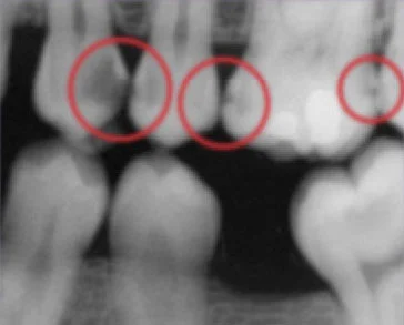
This bitewing shows several areas of decay between (and within) teeth.

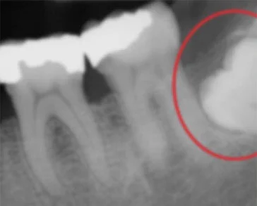
This periapical x-ray shows the top of a molar unable to reach the surface (impacted).

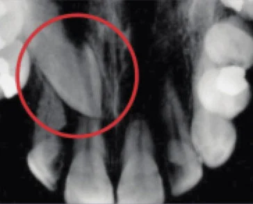
This occlusal x-ray shows a tooth that hasnot yet reached the surface (unerupted).

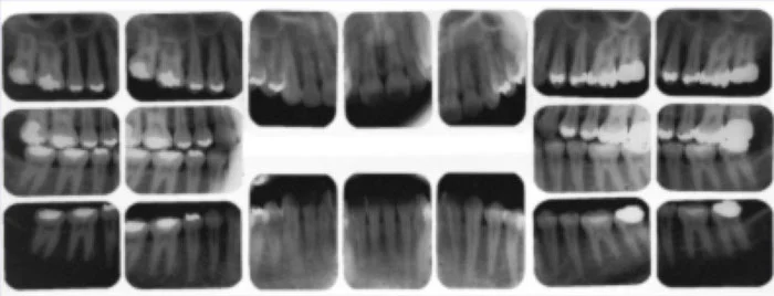
Complete series x-ray.

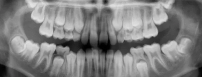
This panoramic x-ray shows baby teeth as well as the developing permanent teeth that have not yet reached the surface.

Ready for a
Healthier Smile?

Office hours

Monday – Thursday: 8:00 AM – 5:00 PM
Friday – Saturday: 8:00 AM – 3:30 PM

Contact

Phone: (775) 825-6655
Fax: (775) 825-8691

Limited Time Offer
Invisalign Special
$4,997
$4,497
Save $500

Fill out the form below to claim your exclusive online discount. Our team will reach out to schedule your consultation.

"*" indicates required fields

MM slash DD slash YYYY

By submitting, you agree to be contacted by White Pine Family Dental. Your information will not be shared.